
Verhoeff-Van Gieson VVG Staining: A Review of Techniques and Applications
Verhoeff-Van Gieson (VVG) staining is a widely used histological technique in medical research and diagnostics. This method involves the use of a specific stain to highlight collagen and elastin in tissue samples, providing valuable insights into tissue structure and disease pathology. With its applications ranging from cancer research to cardiovascular disease diagnosis, VVG staining has become an essential tool in the field of pathology. In this article, we will delve into the context, details, and implications of VVG staining, exploring its common mistakes, smarter alternatives, and future directions.
Context and Background
VVG staining has been a cornerstone of histological analysis for decades, with its origins dating back to the early 20th century. The technique involves the use of a combination of stains, including Verhoeff's elastic stain and Van Gieson's collagen stain, to differentiate between collagen and elastin in tissue samples. This allows researchers and clinicians to visualize the structure and organization of tissues, which is critical in understanding disease mechanisms and developing effective treatments. 
Technique and Applications
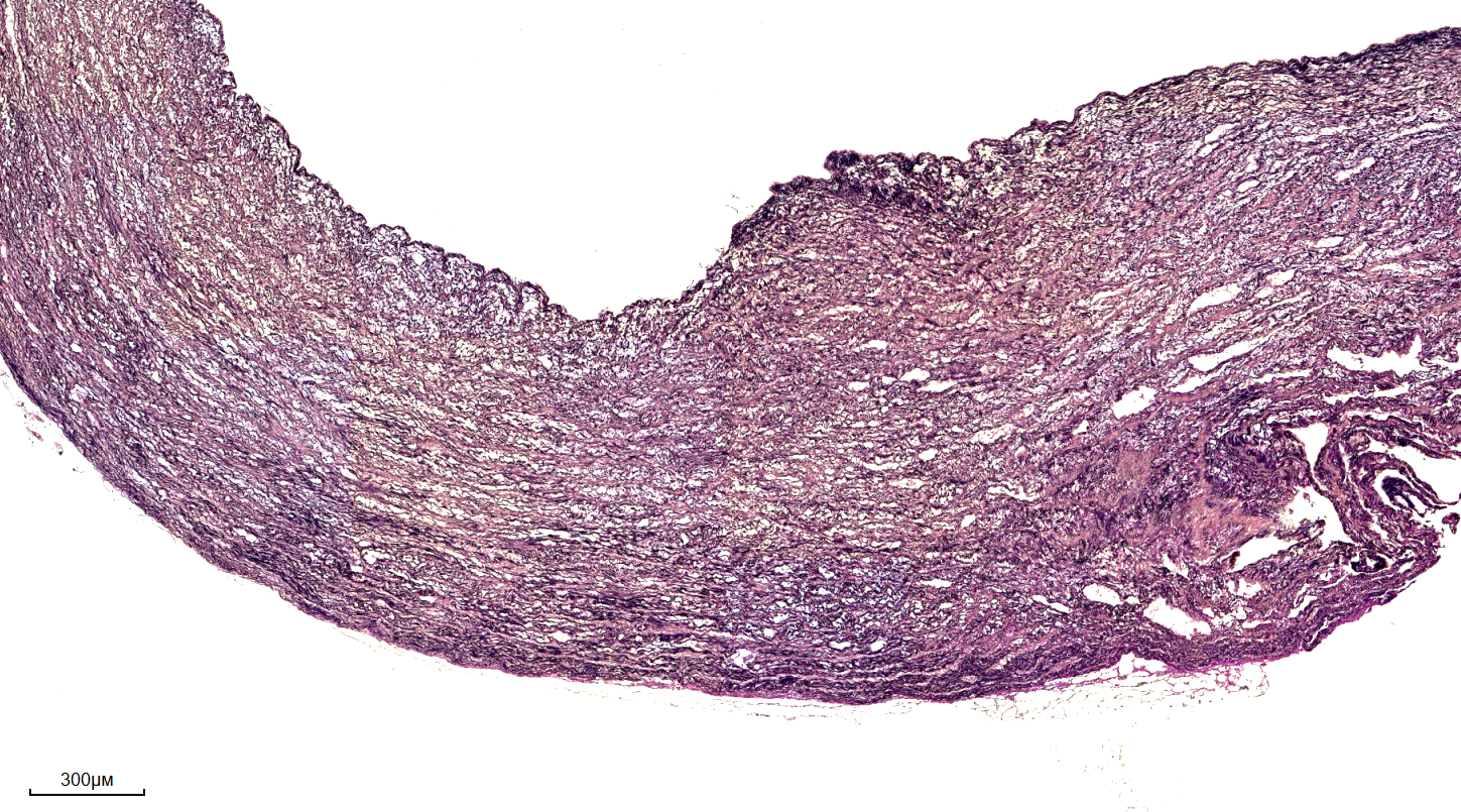
The VVG staining technique involves a series of steps, including tissue fixation, sectioning, and staining. The resulting stained sections can be visualized using light microscopy, providing detailed information on tissue structure and composition. VVG staining has a wide range of applications, including cancer research, cardiovascular disease diagnosis, and neurological disorder studies. For example, VVG staining can be used to visualize the structure of blood vessels and identify areas of vascular damage or disease. 
Common Mistakes and Smarter Alternatives
Despite its widespread use, VVG staining is not without its limitations and potential pitfalls. One common mistake is the use of incorrect staining protocols or inadequate tissue fixation, which can lead to inconsistent or inaccurate results. Additionally, VVG staining can be time-consuming and labor-intensive, requiring specialized expertise and equipment. To address these challenges, researchers and clinicians are exploring alternative staining techniques, such as immunohistochemistry and fluorescence microscopy. These newer methods offer improved sensitivity and specificity, as well as faster turnaround times and reduced labor requirements. Furthermore, the development of digital pathology and artificial intelligence-powered image analysis tools is revolutionizing the field of histology, enabling faster and more accurate analysis of tissue samples.
Implications and Future Directions
In conclusion, VVG staining remains a vital tool in the field of pathology, with its applications continuing to expand and evolve. As researchers and clinicians, it is essential to be aware of the potential pitfalls and limitations of this technique, as well as the smarter alternatives and emerging technologies that are transforming the field of histology. By embracing these advances and adopting best practices, we can improve our understanding of disease mechanisms, develop more effective treatments, and ultimately enhance patient outcomes. As the field of pathology continues to evolve, it is likely that VVG staining will remain a cornerstone of histological analysis, with its applications extending into new and exciting areas of research and diagnostics.
Verhoeff-van Gieson (VVG) Histology And Analysis. VVG-stained Cross

Verhoeff-van Gieson (VVG) histology and analysis. VVG-stained cross ...
Verhoeff-Van Gieson Stain(VVG)_規格表 - BioTnA
Verhoeff-Van Gieson stain(VVG)_規格表 - BioTnA
Verhoeff-van Gieson (VVG) Histology And Analysis. VVG-stained Cross
Verhoeff-van Gieson (VVG) histology and analysis. VVG-stained cross ...
Verhoeff-Van Gieson (VVG) Staining Protocol For Elastic Fibers - IHC WORLD

Verhoeff-Van Gieson (VVG) Staining Protocol for Elastic Fibers - IHC WORLD